
Skeletal Muscle vs Smooth Muscle vs Cardiac Muscle
Human bodies rely on specialized muscle tissues to generate physical force and produce movement. Three distinct muscle types exist within the human body: skeletal muscle, smooth muscle, and cardiac muscle.
Each muscle type performs specific structural, functional, and physiological roles necessary for survival. Understanding these three muscle tissue types reveals how human bodies run, digest food, and pump blood continuously. This comprehensive guide breaks down the key biological differences between skeletal, smooth, and cardiac muscle tissues.
Overview of the Three Primary Muscle Tissues
Muscle tissue accounts for a massive portion of total human body mass. Specialized muscle cells contract to produce bodily motion, maintain physical posture, and circulate vital fluids.
While all three muscle types generate physical mechanical force, their structural designs differ significantly. Anatomists categorize these muscle tissues based on appearance, physiological control, and bodily location.
- Skeletal muscle anchors to bones to move limbs and execute voluntary body movements.
- Smooth muscle lines hollow internal organs to move substances through digestive and vascular tracks.
- Cardiac muscle forms the muscular heart wall to pump oxygenated blood throughout the circulatory system.
- All three tissue types require adenosine triphosphate to power mechanical cellular contraction.
Each muscle type adapts perfectly to its specific biological function within the human body. Learning their anatomical features clarifies how human physiology functions during rest and exercise.
1. Structural Architecture and Microscopic Appearance
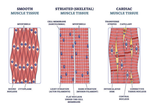
Examining muscle tissue under a microscope reveals distinct structural differences between the three muscle types. Skeletal and cardiac muscles feature visible alternating light and dark bands called striations.
Smooth muscle completely lacks these striated bands, presenting a uniform, smooth appearance under microscopic magnification. The presence or absence of microfilaments determines these unique structural patterns.
| Muscle Type Feature | Skeletal Muscle | Smooth Muscle | Cardiac Muscle |
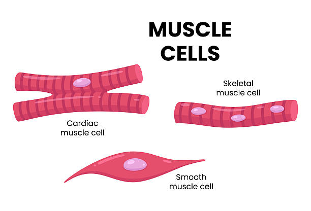
| Cell Shape | Long cylindrical fibers | Spindle-shaped cells | Branched Y-shaped fibers |
| Striation Pattern | Present (Striated) | Absent (Non-striated) | Present (Striated) |
| Nuclei per Cell | Multiple peripheral nuclei | Single central nucleus | One or two central nuclei |
These structural arrangements influence how each muscle type generates tension and withstands physical strain. Microscopic architecture aligns directly with the mechanical functional demands of each tissue.
Voluntary Control versus Involuntary Regulation
Control over muscle contractions depends on how the human nervous system connects to muscle tissues. Skeletal muscle operates under conscious somatic nervous system control, allowing deliberate movement.
Conversely, smooth and cardiac muscles operate involuntarily under the control of the autonomic nervous system. You cannot consciously decide to alter smooth muscle contractions or adjust individual cardiac cycles.
- Somatic motor neurons activate skeletal muscle fibers through conscious brain signals.
- Autonomic sympathetic neurons increase involuntary cardiac and smooth muscle contraction speeds.
- Autonomic parasympathetic neurons slow down cardiac contractions and promote smooth muscle digestion.
- Hormones like adrenaline modulate smooth and cardiac muscle performance during physical stress.
This neural organization ensures essential biological processes continue automatically without requiring constant conscious focus. Involuntary regulation keeps your heart beating and digestive organs working during deep sleep.
2. Location and Distribution Across the Human Body
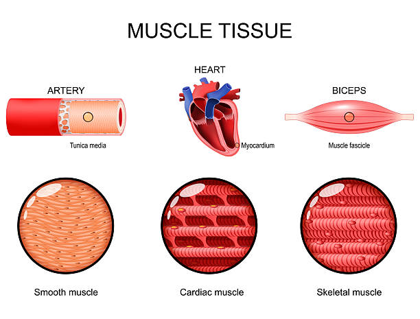
The physical location of each muscle type reflects its primary physiological role within human biology. Skeletal muscle sits distributed across the entire skeleton, attached firmly to bony structures.
Smooth muscle resides primarily inside internal organ walls, blood vessel walls, and microscopic passageways. Cardiac muscle remains strictly localized to a single anatomical organ: the living human heart.
- Skeletal muscles attach to bones via dense connective tissue tendons across every major joint.
- Smooth muscle forms internal layers inside the stomach, intestines, esophagus, and urinary bladder.
- Smooth muscle surrounds arterial walls to regulate arterial lumen diameter and blood pressure.
- Cardiac muscle forms the thick middle layer of the heart wall, known as the myocardium.
Recognizing where these tissues exist helps explain how organ systems coordinate daily physical functions. Location defines how each muscle interacts with surrounding body systems.
Cellular Nuclei Distribution and Structural Geometry
Muscle cells vary dramatically in their internal cellular geometry and nuclear arrangement. Skeletal muscle fibers fuse during embryonic development, creating massive, multi-nucleated cylindrical structures.
Smooth muscle cells retain a compact, spindle-shaped structure with tapered ends and a single nucleus. Cardiac muscle fibers branch intricately, connecting end-to-end to form a dense, interconnected three-dimensional network.
| Cellular Characteristic | Skeletal Muscle Fibers | Smooth Muscle Cells | Cardiac Muscle Fibers |
| Cell Length | Up to 30 centimeters | 20 to 500 micrometers | 50 to 100 micrometers |
| Cell Interconnections | Isolated parallel fibers | Gap junctions in single-unit | Intercalated discs with gap junctions |
| Regenerative Ability | Limited via satellite cells | Moderate cellular division | Extremely minimal repair rate |
Nuclear placement reflects the transport and protein synthesis demands of each specific cell structure. Multi-nucleated skeletal fibers manage protein synthesis across exceptionally long physical distances.
3. Mechanisms of Muscle Contraction and Calcium Signaling
All three muscle types use actin and myosin protein filaments to generate mechanical contraction force. However, the precise molecular signaling pathways that trigger filament sliding vary between muscle tissues.
Skeletal and cardiac muscles rely on calcium binding to troponin to expose active myosin-binding sites. Smooth muscle lacks troponin, utilizing calmodulin and myosin light-chain kinase to initiate contraction.
- Skeletal muscle releases stored calcium rapidly from an extensive sarcoplasmic reticulum network.
- Cardiac muscle relies on extracellular calcium entry to trigger further internal sarcoplasmic calcium release.
- Smooth muscle utilizes slow extracellular calcium influx to activate calmodulin-dependent phosphorylation pathways.
- ATP breakdown provides the necessary chemical energy for cross-bridge cycling in all three types.
These distinct chemical pathways dictate how quickly each muscle type contracts and relaxes. Molecular variations allow each tissue to fulfill its specific speed and endurance requirements.
Intercalated Discs and Electrical Syncytium in Cardiac Muscle
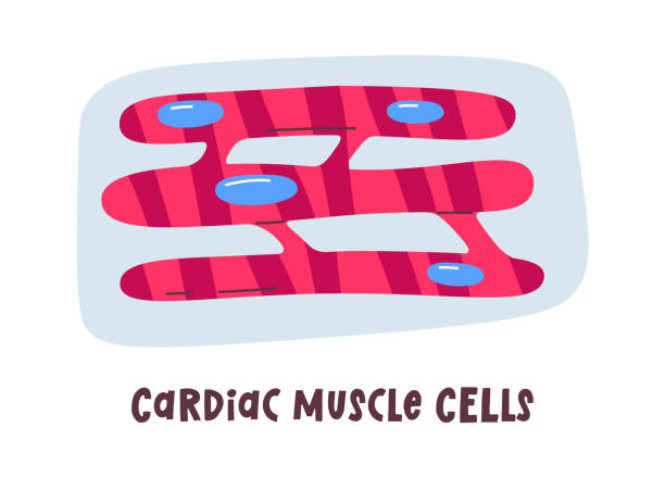
Cardiac muscle possesses unique specialized cellular junctions called intercalated discs that connect adjacent cardiac cells. These structures contain desmosomes for structural anchoring and gap junctions for rapid electrical communication.
Gap junctions allow action potentials to spread almost instantaneously from cell to cell across the myocardium. This electrical continuity causes cardiac muscle fibers to contract together as a single, coordinated functional syncytium.
- Intercalated discs maintain mechanical structural integrity during continuous, high-pressure cardiac pumping cycles.
- Gap junctions permit rapid ion passage, ensuring synchronized chamber contraction across cardiac tissue.
- Desmosomes prevent individual muscle fibers from pulling apart during intense mechanical heart contractions.
- Synchronized contractions maximize blood ejection efficiency from heart chambers into major arteries.
Without intercalated discs, the heart could not pump blood effectively as a unified mechanical unit. This structural feature separates cardiac tissue performance from skeletal and smooth muscle mechanics.
4. Contraction Speed, Force Generation, and Fatigue Resistance
The physical contraction speed and force output of each muscle tissue match its physiological purpose. Skeletal muscle contracts rapidly with immense force, but it exhausts its energy stores quickly during exertion.
Smooth muscle contracts slowly and deliberately, maintaining continuous tonic contractions for hours without fatiguing. Cardiac muscle exhibits rhythmic, moderate-speed contractions, operating continuously without fatigue throughout a person’s lifespan.
| Functional Metric | Skeletal Muscle | Smooth Muscle | Cardiac Muscle |
| Contraction Speed | Fast to exceptionally rapid | Extremely slow and sustained | Moderate rhythmic rate |
| Fatigue Resistance | Low to moderate resistance | High resistance to fatigue | Absolute high fatigue resistance |
| Energy Source | Aerobic and anaerobic pathways | Primarily aerobic metabolism | Strictly aerobic respiration |
These functional differences ensure each muscle type handles its physical workload without failing unexpectedly. Fatigue resistance keeps vital organs functioning continuously around the clock.
Autorhythmycity and Pacemaker Activity
Skeletal muscle requires external nerve impulses from somatic motor neurons to initiate every single contraction cycle. Without nerve stimulation, skeletal muscle remains completely flaccid and paralyzed.
In contrast, cardiac muscle and certain smooth muscles possess intrinsic autorhythmicity, generating their own electrical impulses. Specialized pacemaker cells inside cardiac tissue initiate rhythmic heart contractions independently of external nervous inputs.
- The sinoatrial node acts as the primary natural pacemaker driving rhythmic cardiac contractions.
- Specialized interstitial cells of Cajal generate slow electrical waves inside smooth digestive organs.
- Autonomic nerves alter the rate of intrinsic pacemakers but do not initiate baseline contractions.
- Autorhythmic tissues maintain life-sustaining organ movement even if external neural pathways suffer damage.
Intrinsic rhythmicity ensures continuous cardiac pumping without requiring constant brain signal transmissions. This automatic self-stimulation protects critical life functions against neural interruptions.
5. Blood Supply and Metabolic Demands
Skeletal, smooth, and cardiac muscles demand steady blood flow to supply oxygen and metabolic nutrients. However, cardiac muscle requires the most extensive capillary density due to its absolute reliance on aerobic metabolism.
Cardiac muscle cells pack high concentrations of mitochondria, occupying nearly thirty percent of total cell volume. Skeletal muscle contains variable mitochondrial density based on whether fibers are fast-twitch or slow-twitch types.
- Cardiac tissue extracts up to seventy percent of available oxygen from coronary arterial blood flow.
- Slow-twitch skeletal muscle fibers maintain high capillary density for long-distance endurance activities.
- Fast-twitch skeletal muscle fibers rely on glycogen stores for short, explosive anaerobic efforts.
- Smooth muscle operates on minimal oxygen expenditure, utilizing low metabolic energy rates efficiently.
Metabolic infrastructure matches the daily energy demands placed on each specific tissue type. Rich blood flow prevents metabolic waste accumulation and protects against tissue ischemia.
Elasticity, Extensibility, and The Stress-Relaxation Response
Smooth muscle possesses a unique physiological property known as the stress-relaxation response or plastic adaptation. When a hollow organ like the stomach fills, smooth muscle stretches without increasing internal wall pressure significantly.
This elasticity allows organs to expand and hold variable fluid volumes without triggering premature emptying contractions. Skeletal and cardiac muscles operate on strict length-tension relationships, where overstretching degrades contraction force output.
- Smooth muscle stretches significantly while maintaining consistent baseline structural contraction tension.
- Skeletal muscle generates peak physical force only at optimal baseline muscle resting lengths.
- Cardiac muscle utilizes the Frank-Starling mechanism, increasing contraction force when stretched by returning blood.
- Organ compliance prevents dangerous pressure spikes inside the urinary bladder and gastrointestinal tract.
Specialized elastic properties enable hollow organs to store materials safely until voluntary or involuntary evacuation occurs. Smooth muscle adaptation protects internal organ walls from mechanical rupture under pressure.
6. Muscle Regeneration and Cellular Repair Capacities
When muscle tissues experience physical damage or disease, their ability to repair themselves varies drastically. Skeletal muscle contains specialized satellite cells that divide and fuse to repair damaged skeletal fibers.
Smooth muscle cells retain the highest regenerative capacity, dividing through mitosis to repair injured vascular or organ walls. Cardiac muscle possesses extremely limited regenerative abilities, replacing damaged tissue with non-contractile scar tissue instead.
| Regeneration Metric | Skeletal Muscle | Smooth Muscle | Cardiac Muscle |
| Stem Cell Presence | Satellite cells present | Active cell division capability | Virtually absent stem cells |
| Repair Mechanism | Fiber repair and replacement | Cellular division and hypertrophy | Fibrotic scar tissue formation |
| Functional Recovery | High functional recovery rate | Good tissue repair potential | Permanent functional loss |
Inadequate cardiac muscle regeneration makes heart attacks particularly dangerous to long-term human health. Scar tissue substitution permanently reduces overall cardiac pumping efficiency and mechanical output.
Physiological Roles in Human Homeostasis
The three muscle types work together continuously to maintain internal chemical and physical body homeostasis. Skeletal muscle movement generates internal body heat, helping regulate core body temperature in cold environments.
Smooth muscle adjusts blood vessel diameters to regulate systemic blood pressure and direct blood flow to active tissues. Cardiac muscle maintains continuous perfusion, ensuring every body cell receives fresh oxygen and metabolic nutrients.
- Shivering skeletal muscle contractions convert chemical energy into heat during cold environmental exposure.
- Smooth muscle vasodilation increases blood flow to skin surfaces to release excess internal heat.
- Smooth muscle vasoconstriction redirects warm blood away from skin surfaces to protect core organs.
- Cardiac output adjustments maintain adequate arterial pressure during dramatic physical posture changes.
Every muscle type contributes vital physiological mechanisms that preserve stable internal body conditions. Integrated muscle functions protect the body against environmental and physical stresses.
Neuromuscular Junctions and Synaptic Transmission
Skeletal muscle fibers receive direct innervation through highly structured, individual motor endplates called neuromuscular junctions. Each somatic motor neuron branch forms a precise chemical synapse with a specific skeletal fiber.
Smooth and cardiac muscles feature diffuse neurotransmitter release mechanisms called varicosities or autonomic junctions. Neurotransmitters diffuse over wider physical distances to stimulate multiple smooth or cardiac cells simultaneously.
- Acetylcholine serves as the exclusive neurotransmitter activating skeletal muscle motor endplates directly.
- Acetylcholine and norepinephrine exert opposing regulatory effects on smooth and cardiac muscle cells.
- Synaptic transmission at skeletal motor endplates produces rapid, localized action potential responses.
- Varicosities release neurotransmitters broadly across tissue layers, producing widespread slow tissue responses.
Synaptic architecture determines how precisely the nervous system controls individual muscle fiber groups. Direct neuromuscular endplates enable fine, precise skeletal movements like writing or typing.
Conclusion
Understanding the structural, functional, and physiological differences between skeletal, smooth, and cardiac muscles reveals the brilliant complexity of human anatomy. Skeletal muscle provides voluntary physical movement and strength, allowing you to interact directly with your external environment. Smooth muscle manages involuntary internal transport processes, driving digestion, urinary function, and vascular blood pressure regulation automatically. Cardiac muscle operates continuously as an involuntary, self-stimulating pump, circulating life-giving blood to every cell throughout your lifespan. Each muscle tissue possesses specialized cellular structures, calcium signaling pathways, and energy systems tailored perfectly to its physiological purpose. Appreciating these unique tissue characteristics highlights how seamlessly the human body coordinates movement, maintains homeostasis, and sustains life.
Frequently Asked Questions
Which of the three muscle types is the strongest in the human body?
The answer depends on how you measure strength. The masseter skeletal muscle generates the greatest physical bite force, while the cardiac muscle is the strongest enduring muscle because it pumps continuously without resting.
Can smooth muscle tissue be controlled voluntarily through practice?
No, smooth muscle tissue operates strictly under involuntary autonomic nervous system control. While stress reduction techniques influence autonomic nervous system activity, you cannot consciously contract or relax smooth muscle cells directly.
Why does cardiac muscle not fatigue like skeletal muscle during exercise?
Cardiac muscle contains exceptionally high concentrations of mitochondria and relies strictly on aerobic metabolism. It maintains a constant, rich blood supply through coronary arteries, preventing the accumulation of fatigue-inducing metabolic byproducts.
What happens when cardiac muscle tissue suffers structural damage?
Because cardiac muscle has virtually no regenerative stem cells, dead cardiac cells are replaced by non-contractile fibrotic scar tissue. This scar tissue weakens the heart wall and permanently reduces overall heart pumping efficiency.
How do skeletal muscle fibers increase in size from weightlifting?
Skeletal muscle fibers undergo hypertrophy, expanding in physical diameter by synthesizing additional actin and myosin protein filaments. Existing fibers grow thicker and stronger, rather than dividing to create brand-new muscle cells.
Are all skeletal muscle fibers identical in their performance capabilities?
No, skeletal muscle contains fast-twitch fibers designed for explosive, short-duration power and slow-twitch fibers built for sustained endurance. The proportion of these fiber types varies between different muscles and individual people.
Why is smooth muscle called smooth under a microscope?
Smooth muscle lacks the organized, repeating sarcomere arrangements that create visible light and dark striations in skeletal and cardiac muscles. Under microscopic magnification, smooth muscle cells present a completely smooth, uniform appearance.
Discover more from Muscle Archive
Subscribe to get the latest posts sent to your email.